“You can’t manage what you can’t measure.”
- Peter Drucker
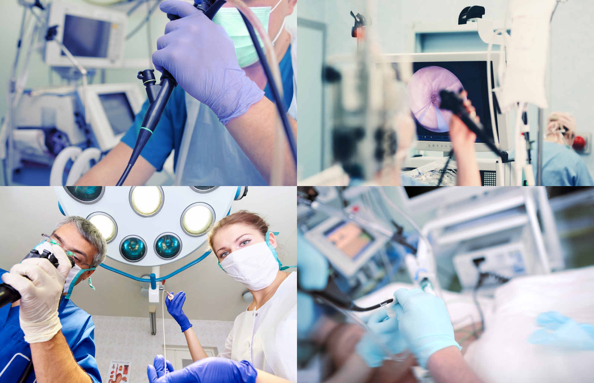
It’s no secret that diagnostic results are the main objective of a navigational bronchoscopy procedure. Early and timely diagnosis can lead to effective treatment and even a cure. Therefore, understanding the exact cause of diagnostic failure is equally as important.
Historically, navigation systems have been evaluated by two criteria: diagnostic yield and localization accuracy. The definition of diagnostic yield varies substantially across clinical studies but conservatively it is defined as the likelihood to obtain a definitive histologic diagnosis (malignant or benign) on the day of the procedure. Localization is defined as the ability to locate an object's position through the processing and interpretation of indirect information provided by means of nonvisual and virtual sources. A well-known example of a localization technique is the triangulation method. This method is based on the received signal strength from several signal transmitters with a known location. These techniques, collectively called localization methods, may use electromagnetic sensors, sensor arrays, shape sensors, laser radars, navigation sensors, etc. as a basic source of information for calculation.
By definition, localization--as any computational technique--involves inaccuracy. For instance, the accuracy of a car’s GPS may vary between 2 to 10 meters, depending on the calculation technique. Localized objects can also be visualized or rendered (drawn on a computer screen) through computer graphic techniques, creating an illusion of display in the real world. When referencing localization, it’s imperative to recognize the difference between its virtual visualization, which can be inaccurate, and actual sight of the object in the real world.1
Several pulmonology studies have revealed an unexplained gap between high localization and low diagnostic yield. For example, studies have shown that there is an unexplained gap between radial endobronchial ultrasound (rEBUS) localization (reported as more than 90%) vs. diagnostic yield (reported between 43% to 57%), when rEBUS is used alone or in combination with electromagnetic navigation bronchoscopy (ENB).2,3 The Gex8 meta-analysis of ENB demonstrates a “localization success” of 97.4% but only had a diagnostic yield of 64.9%.
Dr. Gildea has further elaborated on this concept in his retrospective analysis, “We are beginning to understand that all advanced bronchoscopy techniques used to sample peripheral lesions have significant limitations, as noted by the drop-off between lesion localization and biopsy diagnostic yield, even when operator expertise is assured in procedural, navigation, and real-time localization.” 4
Even when localization is perceived as “real-time” it fails to show the real-time location of a lung lesion relative to the biopsy tool. Researchers are forced to hypothesize as to the root cause for this unexplained gap, rather than making evidence-based conclusions.
Computed tomography (CT) guided transthoracic needle aspiration (TTNA) has demonstrated a high diagnostic yield of 83.7 % for pulmonary lesions of less than 15 mm and 96.8 % for lesions larger than 15 mm.7
TTNA involves using computed tomography (CT) imaging to see the biopsy needle in the lesion before taking the tissue samples. This “tool-in-lesion” confirmation is certainly the crucial step associated with the high diagnostic yield of this procedure. However, CT-guided TTNA is associated with a higher rate of complications, mainly pneumothorax, with a reported pooled rate of 25.9% as well as pulmonary hemorrhage. 5-7
Between advanced bronchoscopy techniques like electromagnetic navigation, robotic bronchoscopy, or rEBUS, and the clinical constraints of TTNA, a new approach was necessary. Such an approach would have to combine the “low patient risk” profile of navigational bronchoscopy with the diagnostic success and consistency of TTNA.
In 2014, Body Vision was founded to provide physicians a level of control and confidence that exists in TTNA, during navigational bronchoscopy. The Body Vision platform is revolutionizing advanced bronchoscopy techniques by replacing inaccurate localization concepts with advanced imaging and real-time, tool-in-lesion confirmation. This easy access to advanced capabilities is possible without the use of electromagnetic, shape, or ultrasonic sensors. Through C-arm based tomography (CABT), our technology offers accurate, real-time imaging with any conventional C-Arm. CABT provides 3D images of the biopsy tool and lesion, during the procedure, similar to preoperative CT. The ability to see the location and orientation of the tool and lesion before biopsy tissue sampling assures that samples will be collected from the desired location of the suspicious lesion.
“Advanced CABT imaging allows physicians to manage the procedure with confidence,” says Dr. Michael Pritchett, Pulmonologist at Pinehurst Medical Clinic and Director of the Chest Center of the Carolinas at FirstHealth MRH. “Body Vision technology gives you the ability to see your tool or catheter with respect to the lesion, which in turn can help the clinician make the adjustments necessary to be on target.”
In summary, an advantage of Body Vision’s CABT is its ability to assist physicians in identifying, analyzing and addressing the root cause of a failed procedure. Failure can be due to the morphology of the lesion, the samples collected, or the sampling strategy. The Body Vision platform eliminates this question of accuracy by providing doctors with the ultimate control and enabling them to get real-time imaging information during the procedure vs. making an intelligent guess.
Within advanced bronchoscopy, we are often asked “What is your diagnostic yield?” Without neglecting this important question, may I suggest rephrasing the famous statement by Peter Drucker, “You are likely to improve what you manage. You are capable of correcting mistakes that you are able to see and be aware of.”
When considering an advanced guided bronchoscopy platform, it’s important to recognize that the platform by itself is not a magical solution for the problem of diagnostic yield. In the hands of the experienced bronchoscopist, a guided navigation platform is an opportunity to improve clinical outcomes. Therefore, the right question to be asking is: “Does this platform provide the level of confidence sufficient to analyze and help to resolve any potential diagnostic failures?”
Body Vision has reimagined navigational bronchoscopy and made advanced real-time imaging available for every bronchoscopy suite as the basis of the next generation of guided bronchoscopy platforms. As we look to the future, we believe in creating a managed, evidence-based approach that gives physicians a superior level of confidence and satisfaction.
References:
1. Pritchett MA, Bhadra K, Calcutt M, Folch E. Virtual or reality: divergence between preprocedural computed tomography scans and lung anatomy during guided bronchoscopy. J Thorac Dis. 2020 Apr;12(4):1595-1611
2. Chen A, Stather DR, Maceachern P, et al. Diagnostic utility of peripheral endobronchial ultrasound with electromagnetic navigation bronchoscopy in peripheral lung nodules. Respirology. 2013;18(5):784-789. doi:10.1111/resp.12085
3. Chen AC, Loiselle A, Zhou L, Baty J, Misselhorn D. Localization of Peripheral Pulmonary Lesions Using a Method of Computed Tomography–Anatomic Correlation and Radial Probe Endobronchial Ultrasound Confirmation. Ann Am Thorac Soc. 2016 Sep;13(9):1586-92.
4. Gildea TR. Lung Lesion Localization and the Diagnostic Drop. Annals of the American Thoracic Society. 2016;13(9):1450-1452. doi:10.1513/annalsats.201606-509ed
5. DiBardino DM, Yarmus LB, Semaan RW. Transthoracic needle biopsy of the lung. Journal of Thoracic Disease. 2015;4. doi:10.3978/j.issn.2072-1439.2015.12.16
6. Huo YR, Chan MV, Habib A, Lui I, Ridley L. Pneumothorax rates in CT-Guided lung biopsies: a comprehensive systematic review and meta-analysis of risk factors. Br J Radiol. 2020 Apr
7. Huang MD, Weng HH, Hsu SL, Hsu LS,1 Lin WM, Chen CW, Tsai YH. Accuracy and complications of CT-guided pulmonary core biopsy in small nodules: a single-center experience. Cancer Imaging. 2019; 19: 51.
8. Gregoire Gex 1, Jacques A Pralong, Christophe Combescure, Luis Seijo, Thierry Rochat, Paola M Soccal,. Diagnostic yield and safety of electromagnetic navigation bronchoscopy for lung nodules: a systematic review and meta-analysis, Respiration, 2014; 87(2):165-76.